



Blood Clot in the Brain: A Guide to Causes, Symptoms, and Treatment

Searching for information about a "blood clot in the brain" often leads to a mix of confusing medical jargon, frightening stories, and conflicting advice. If you're feeling overwhelmed, that's understandable. This is a common and frustrating position when all you need is a clear, reliable explanation.
The goal of this article is to provide that clarity. We will explain how healthcare professionals understand this serious condition, from the first warning signs to the path of recovery. This is not about miracle claims or false promises, but about building a solid understanding of what a blood clot in the brain is, what it means, and how it is managed.
This visual guide shows the path we will follow, moving from initial confusion toward a clearer perspective.
As this illustrates, a structured approach to a complex health topic is often the most effective way to feel more in control and better understand the situation.
The Two Main Types of Blood Clots in the Brain
When clinicians discuss a blood clot in the brain, they are usually referring to one of two distinct events. Understanding this difference is a crucial first step.
The key distinction lies in where the clot is located: in an artery that carries blood to the brain, or in a vein that drains blood away from it.
Two Main Types of Blood Clots in the Brain
| Feature | Ischaemic Stroke (Clot in an Artery) | Cerebral Venous Sinus Thrombosis (CVST) (Clot in a Vein) |
|---|---|---|
| Where the Clot Forms | In an artery, one of the vessels delivering oxygen-rich blood to the brain. | In a venous sinus, which are large veins responsible for draining used blood from the brain. |
| The Primary Problem | Interruption of supply. The clot prevents oxygen and nutrients from reaching a part of the brain, causing tissue damage. | Drainage backup. The clot creates a "traffic jam" of blood trying to exit the brain, which leads to swelling and increased pressure. |
| How Common Is It? | Very common. This is the cause of approximately 85% of all strokes. | Very rare. CVST is significantly less common than ischaemic stroke. |
| Analogy | Like a blocked pipe leading into a house, cutting off the water supply. | Like a blocked drain that causes water to back up and flood the house. |
| Immediate Goal of Treatment | To restore blood flow as quickly as possible, often by dissolving or removing the clot. | To prevent the clot from enlarging and to reduce pressure inside the head, typically using blood thinners. |
As the table shows, although both conditions involve a blood clot, the underlying mechanics are entirely different. This is why a rapid and accurate diagnosis is so critical for the medical team.
In a clinical setting, the first priority is to determine whether the clot is in an artery or a vein. This single piece of information dictates the entire treatment strategy.
While the sudden vascular nature of a brain clot is unique, the brain's response can sometimes share similarities with other neurological issues. You can explore how different brain injuries are managed by reading about the long-term challenges of post-concussion syndrome.
By understanding these foundational concepts, you can feel more prepared and less intimidated when speaking with healthcare professionals, whether for yourself or for someone you care about.
How a Blood Clot Disrupts the Brain
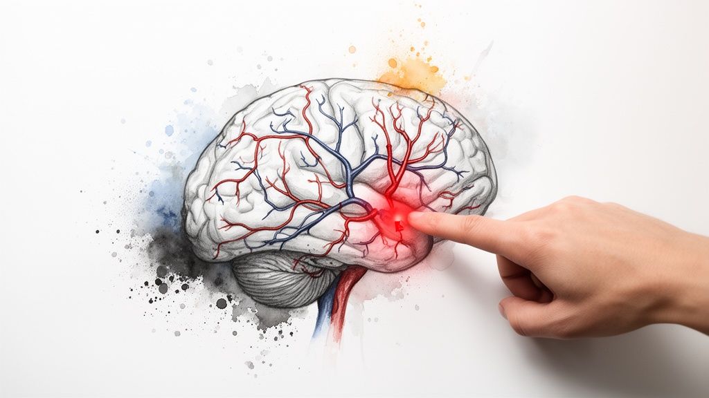
To fully appreciate why a blood clot in the brain is a medical emergency, it helps to view the brain's blood supply as a continuous delivery network where traffic must always keep moving.
Arteries are the highways bringing oxygenated blood to brain cells, delivering essential fuel. Veins are the routes that carry used blood and waste products away. For the brain to function correctly, both pathways must remain clear.
A blood clot disrupts this system in one of two ways, depending on its location.
When a Supply Artery Is Blocked
This is what occurs during an ischaemic stroke, which accounts for about 85% of all strokes. A clot becomes lodged in an artery, acting like a sudden roadblock on a vital supply route.
The moment this happens, blood flow beyond the blockage ceases. The area of the brain that the artery supplied is immediately cut off from oxygen and nutrients.
Brain cells can begin to die within minutes of being deprived of blood. This is why an ischaemic stroke is a time-critical emergency where every second counts toward preserving brain function.
The specific symptoms that appear—such as one-sided weakness, slurred speech, or facial drooping—are a direct result of which part of the brain is being starved of blood.
When a Drainage Vein Is Blocked
A much rarer but equally serious event is a Cerebral Venous Sinus Thrombosis (CVST). This occurs when a clot forms in one of the brain's venous sinuses—the large veins that drain blood away from the brain.
This is like a blocked drain. Blood cannot leave the brain as it should, resulting in a dangerous backup.
This congestion leads to a rapid increase in pressure inside the skull, which can trigger several serious problems:
- Brain Swelling (Cerebral Edema): The brain may begin to swell, but it has no room to expand within the rigid skull.
- Bleeding (Haemorrhage): The intense pressure can cause fragile blood vessels to rupture, leading to a bleed in the brain.
Unlike an ischaemic stroke with its localised symptoms, the signs of CVST are often more general. They can include a severe, persistent headache, blurred vision, or seizures. This is because the problem is a generalised pressure increase across the whole brain, not damage to a specific area.
The mental cloudiness that can follow such events is often described as 'brain fog'. For a deeper understanding of this symptom, you can learn more about what causes brain fog symptoms in our related article.
Whether the clot blocks a supply route or a drainage path, the result is a sudden crisis in the brain's balanced environment, leading to immediate and often significant symptoms.
Recognising Critical Symptoms and When to Act

When dealing with a potential blood clot in the brain, recognising the symptoms is the single most important factor for improving outcomes. Healthcare professionals often say "time is brain" because every second that passes can affect recovery.
Clinicians are trained to identify key patterns in an emergency. One of the most effective tools for the public to identify a potential ischaemic stroke is the F.A.S.T. test. It is a simple checklist anyone can use.
Using the F.A.S.T. Test
The strength of the F.A.S.T. acronym is its simplicity. It breaks down the most common physical signs of a stroke into three quick observations, followed by an urgent action.
-
F – Face Drooping: Ask the person to smile. Does one side of their face sag or seem numb? An uneven smile can be a classic sign that the part of the brain controlling facial muscles is affected.
-
A – Arm Weakness: Ask them to raise both arms. Does one arm drift downwards? They might struggle to lift one arm as high as the other. This suggests weakness on one side of thebody.
-
S – Speech Difficulty: Ask the person to repeat a simple phrase, like "the sky is blue." Is their speech slurred or unusual? Are they unable to find the words? Sometimes, they may not be able to speak at all.
T – Time to call 999: If you notice any of these signs, even if they seem to resolve, it is time to call 999 for an ambulance immediately. It is also very helpful to note the time the first symptoms appeared, as this information is vital for the medical team.
This simple test is an essential first-response tool. Public awareness of these signs is crucial.
Symptoms Beyond the F.A.S.T. Model
While F.A.S.T. is effective for identifying common stroke patterns, a blood clot in the brain can present in other ways. This is particularly true for rarer conditions like Cerebral Venous Sinus Thrombosis (CVST).
One major red flag that clinicians always look for is a sudden, severe headache. Patients often describe it as the "worst headache of my life." This is typically not like a normal migraine; it tends to be abrupt and intense, often caused by the pressure build-up from a venous clot.
Other critical symptoms to be aware of include:
- Sudden Confusion: An abrupt change in mental state, such as difficulty thinking clearly or understanding what is happening.
- Vision Problems: Sudden blurred or double vision, or a loss of sight in one or both eyes.
- Loss of Balance or Coordination: A sudden difficulty with walking, dizziness, or feeling unsteady.
Recognising these signs is the first step. The challenges that follow brain-related events are explored in our article on what post-concussion syndrome is, which offers more insight into the brain's response to injury.
Regardless of the specific sign, the message is consistent: any sudden, severe, and unexplained neurological symptom requires immediate medical attention. Do not wait to see if it improves. Call for help.
How Clinicians Diagnose a Blood Clot in the Brain
When a person arrives at a hospital with suspected stroke symptoms, the medical team begins a rapid and coordinated process. The first priority is to determine exactly what is happening inside the brain. This involves a sequence of steps designed to build a complete picture quickly.
The investigation almost always starts with brain imaging. Clinicians need to see inside the head to understand the problem, and they need to do so without delay.
The First Step: Brain Imaging
The standard initial test is a CT (Computed Tomography) scan. This is like a sophisticated X-ray that creates detailed cross-sectional images of the brain. The main advantage of a CT scan is its speed; it can be completed in minutes, which is critical in an emergency.
The primary purpose of this first scan is to check for bleeding. If a bleed is present (a haemorrhagic stroke), then clot-busting medications are not an option. Differentiating between a clot (ischaemic stroke) and a bleed is the first and most crucial decision point for treatment.
Following the CT scan, an MRI (Magnetic Resonance Imaging) scan may be performed. An MRI provides a more detailed view of the brain’s soft tissues. It is particularly good at pinpointing the exact location and extent of damage caused by a clot, which helps to confirm an ischaemic stroke diagnosis.
Mapping the Brain's Blood Vessels
To get an even closer look at the blood vessels, clinicians often use specialised imaging techniques. These tests create a "road map" of the brain's arteries and veins, showing exactly where a blockage is.
- CTA (CT Angiography): This involves injecting a contrast dye into a vein before the CT scan. The dye illuminates the blood vessels on the scan, making any clots more visible.
- MRA (MR Angiography): This works on a similar principle but uses an MRI scanner and can often be done without a contrast dye injection.
These scans are vital for confirming conditions like Cerebral Venous Sinus Thrombosis (CVST) and for planning interventions, such as a thrombectomy, where the clot is physically removed.
In clinical settings, the choice between CT and MRI often comes down to speed versus detail. CT provides the quick initial look needed to rule out bleeding. MRI offers the in-depth analysis required to guide longer-term treatment and prognosis.
Completing the Picture with Other Tests
In addition to imaging, other tests help clinicians understand the "why" behind the clot and assess the person's overall health. Blood tests are standard practice, checking for factors like blood sugar levels, kidney function, and blood clotting times.
This proactive approach to risk is becoming standard practice. For example, the NHS England's VTE prevention programme highlights the importance of assessing patients for venous thromboembolism (VTE) risk upon hospital admission. Systematic screening is a crucial part of preventing clots, including those that can affect the brain.
By combining information from rapid brain scans, detailed vessel mapping, and supportive blood tests, the medical team can confidently diagnose a blood clot in the brain, determine its type, and initiate the most effective treatment promptly.
Treatment Approaches and The Recovery Journey

When a blood clot in the brain is diagnosed, the medical response is swift and tailored to the situation. The specific approach depends on the type of clot, its location, and the patient's overall health.
For an ischaemic stroke (a blocked artery), the primary goal is to restore blood flow to the affected brain area as quickly as possible. This is commonly addressed with one of two key emergency treatments.
Emergency Treatments for Ischaemic Stroke
The first-line treatment is often a medication-based therapy called thrombolysis. This involves administering powerful "clot-busting" drugs through an IV drip. These drugs work to dissolve the blockage and reopen the artery. However, this treatment is time-sensitive and must typically be given within a few hours of the first symptoms.
If a patient is not eligible for thrombolysis, or if the clot is large, clinicians may perform a procedure called a thrombectomy. In this procedure, a specialist guides a catheter through blood vessels to the clot in the brain. A small device is then used to physically retrieve the clot, immediately restoring blood flow.
Managing Cerebral Venous Sinus Thrombosis (CVST)
Treating a CVST involves a different strategy. Here, the problem is a blockage in the brain's drainage system. The primary treatment is anticoagulant medications, also known as blood thinners.
These drugs do not dissolve the existing clot directly. Instead, they prevent the clot from growing larger and stop new ones from forming. This allows the body's natural clot-dissolving mechanisms to gradually break down the blockage over time. This treatment is often required for several months.
The Gradual Process of Rehabilitation
Once the immediate medical crisis is over, the journey of recovery begins. This is a gradual and supportive process focused on helping the brain to heal and adapt. Rehabilitation is a collaborative effort involving a team of specialists.
The core principle of rehabilitation is neuroplasticity—the brain’s remarkable ability to reorganise itself by forming new neural connections. Each therapy session is designed to stimulate this process, encouraging healthy parts of the brain to take over functions from damaged areas.
This multidisciplinary team often includes:
- Physiotherapists, who help with regaining movement, strength, balance, and coordination.
- Occupational Therapists, who focus on relearning daily activities like dressing or preparing meals to restore independence.
- Speech and Language Therapists, who assist with communication difficulties (aphasia) or swallowing problems (dysphagia).
For many, specialised after stroke care is a vital part of regaining function and improving quality of life.
The recovery process varies for each individual. Progress can be slow at times, with periods of advancement and plateaus. The challenges are not just physical but can also be emotional and cognitive. The goal is to support each person in adapting to any lasting changes and achieving the best possible outcome.
Frequently Asked Questions About Brain Clots
It is natural to have many questions about a serious topic like a blood clot in the brain. Here are clear answers to some of the most common ones.
Can a Blood Clot in the Brain Heal on Its Own?
Waiting for a brain clot to resolve on its own is extremely dangerous. During an ischaemic stroke, brain tissue is being deprived of oxygen, and damage can become irreversible within minutes.
While the body has natural systems for breaking down clots, this process is far too slow to be effective in an emergency like a stroke. The purpose of acute treatment is to restore blood flow as quickly as possible. In the case of a CVST, anticoagulant medications are essential to prevent the clot from worsening. Any suspected brain clot requires immediate medical evaluation.
What Is the Difference Between an Aneurysm and a Blood Clot?
It is common to confuse these two conditions, but they are fundamentally different problems, even though both can lead to a stroke.
- A blood clot is a blockage inside a blood vessel that stops blood from passing through. This causes an ischaemic stroke.
- An aneurysm is a weak, balloon-like bulge on the wall of a blood vessel. The primary danger is that it can rupture, causing blood to leak into the brain. This causes a haemorrhagic stroke, or a "bleed."
So, one is a blockage, and the other is a bleed. An emergency CT scan is the first step in telling them apart, as their treatments are completely different.
In the hospital, the first question is always "clot or bleed?" A clot starves the brain of blood. A bleed floods it, causing damage from pressure and irritation. The symptoms can look very similar, but what's happening inside the head—and what we need to do about it—couldn't be more different.
Are There Warning Signs Before a Major Brain Clot Occurs?
Sometimes, yes. The most significant warning sign is a Transient Ischaemic Attack (TIA), often called a "mini-stroke." This happens when a clot temporarily blocks a blood vessel, causing stroke-like symptoms that usually disappear within an hour.
The symptoms of a TIA are the same as a major stroke—the F.A.S.T. signs apply. A TIA should never be ignored. It is a major red flag that a more serious, permanent stroke may be imminent. It serves as a critical warning, providing a window of opportunity to seek medical attention, determine the cause, and start treatment to prevent a worse outcome.
Can Stress Cause a Blood Clot in the Brain?
While stress is not a direct cause of a blood clot in the brain, chronic stress can contribute to the underlying risk factors.
Sustained stress can elevate blood pressure, increase inflammation, and may lead to lifestyle habits that are harmful to cardiovascular health, such as a poor diet or lack of exercise. These are all factors that can increase the risk of clot formation over time.
The more direct causes of a blood clot in the brain are typically conditions like atrial fibrillation (an irregular heartbeat), high cholesterol, and atherosclerosis (hardening of the arteries). So, while clinicians would not say stress causes a stroke, managing it is an important part of maintaining cardiovascular health and lowering overall risk.
This article aims to reduce confusion and provide a clearer understanding of a complex topic. However, blog posts have their limits. For readers who want to go beyond articles and get a more structured overview of how to manage their health, The Patients Guide was created.
Our guides offer a deeper, more organised way to connect symptoms, causes, and treatments. They are designed for people who want clarity that lasts.
Explore all available guides at https://www.thepatientsguide.co.uk.
Also in News


Blood Test Results Time: Your UK Guide
Get precise blood test results time info for the UK in 2026. Understand NHS turnaround, common delays, how to check your results, and when to contact your GP.
Read More


Headache Quick Remedy: Immediate Relief Guide
Headache quick remedy - Find a headache quick remedy that works. Our guide helps you understand your headache and choose safe, effective action for immediate
Read More


Mouth Ulcers Treatment: A Clear Guide
A calm, clear guide to mouth ulcers treatment. Learn about causes, self-care, OTC options, when to see a doctor, and how to prevent recurring ulcers.
Read More